Studies show that early detection of breast cancer is the most important factor in determining an effective course of treatment. In fact, finding breast cancer early, before it has a chance to spread, increases the five-year survival rate of women with stage 0 or stage I breast cancer to almost 100%.
According to BreastCancer.org, one in eight women in the U.S. will develop invasive breast cancer throughout the course of her lifetime, which is why scheduling a regular breast exam is so important. Medical scientists have made great strides in developing more effective breast-screening techniques - like digital breast tomosynthesis (also known as 3-D mammogram) - and taking advantage of that technology can be potentially life-saving.
What is a 3-D Mammogram?
Digital breast tomosynthesis (3-D mammogram/tomosynthesis) is an, FDA-approved imaging technology that helps physicians detect smaller tumors at the earliest stages of breast cancer. While the procedure is similar to a standard mammogram, tomosynthesis enables radiologists to see inside the breast in greater detail.
By taking several low-dose X-ray images from different angles, an imaging technician can combine these tiny pictures (each about one millimeter in size) and generate a composite 3-D picture of the breast. This greatly reduces or eliminates problems found in traditional mammography (like ‘false positives’), that can be caused when dense breast tissue overlaps.
3-D Mammograms vs. 2-D Mammograms
Though 2-D and 3-D mammograms essentially perform the same function - examining the breast for cancer - the two procedures are quite different. For example, think of the contrasts between a standard X-ray and a CT Scan. While both procedures can look at the same parts of the body, the amount of information they provide is vastly different.
A standard mammogram takes two-dimensional images of the breast - one from the top and one from the side. However, the human body is not two-dimensional, and when converted to two flat images, a lot of information can be lost.
On the other hand, a 3-D mammogram utilizes a mobile X-ray unit that moves around the breast, capturing a succession of images that are assembled digitally into a three-dimensional model. This means the radiologist can evaluate a more complete image of the inside of the breast, including any small findings that may not be visible on a conventional 2-D mammogram.
Ideal Procedure for Dense Breast Tissue
Another benefit of a 3-D mammogram is its enhanced imaging for women with dense breast tissue. If you’ve already had a breast cancer screening, you’ve likely discussed your breast tissue status with your physician.
Breasts are considered 'dense' if they have an overabundance of fibrous or glandular tissue and minimal amounts of fat. Approximately 43% of women, between the ages of 40 and 74, are classified as having dense breasts. It can be more difficult for radiologists reviewing traditional mammograms to detect breast cancer in patients with dense breast tissue, and research shows that women with dense breasts are six times more likely to develop breast cancer.
In 2011, the Texas Legislature passed a law, often referred to as Henda's Law, which requires a mammography facility to notify all women with dense breast tissue that traditional mammograms could hide abnormalities and other risk factors for breast cancer, and that they could benefit from women "supplemental" screening techniques, such as breast ultrasounds and 3-D mammograms, both of which have been proven to detect more cancer in dense breast tissue If you have dense breasts, ask your physician about the benefits of additional imaging procedures, like 3-D mammograms.
Compression is Key to Better Images
It's recommended that women should schedule regular breast cancer screenings starting at age 40, but many women put off the procedure because mammograms can be a bit uncomfortable.
During a standard 2-D mammogram, the breast is positioned on a detector plate and compressed with a plastic paddle to ensure a good diagnostic image. This is necessary to make sure the whole breast is scanned. (Remember, normal mammograms take flat, two-dimensional images of the top and side of each breast.)
A 3-D mammogram requires the same amount of compression as a Full Field Digital Mammography. Having optimal compression is important, especially during a 3-D mammogram. The X-ray tube moves across the breast, taking many small images.

Mammogram Reminder
Early detection is the best defense against breast cancer, which is why Memorial Hermann can send you a reminder to schedule your annual screening mammogram.
Remind MeWhat to Expect From a 3-D Mammogram
To prepare for your 3-D mammogram:
- Consider scheduling your breast exam one to two weeks after your period begins. Breasts are the least tender just after menstruation, which will make the exam more comfortable.
- Wear loose-fitting, easy-to-remove clothing. You will be asked to strip to the waist for the exam and remove all jewelry.
- Do not apply deodorant, perfume, lotion, or other products to your underarms or torso before the exam. These items can sometimes appear as 'false positives' on scans.
- Inform your physician if you are pregnant, breastfeeding, have breast implants or are currently suffering from any medical conditions before scheduling your exam.
How Does a 3-D Mammogram Work?
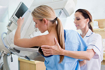
Your breast exam will be conducted in a private room by a radiologic technologist. The technologist will have you stand in front of the 3-D mammography machine, and your breast will be positioned on a detector plate. The technologist will then use a plastic compression paddle to compress and hold your breast in place. Any movement can blur the image, so to avoid the need for additional images, it's important to stay as still as possible throughout the exam. If at any point, you're feeling too much discomfort, don't hesitate to let the technologist know.
When you're ready, the technologist will start the 3-D mammography machine. The X-ray tube will move over your breast in an arc, capturing multiple X-ray images as it moves.
Once your exam is complete, the images are sent to a computer to construct a 3-D composite image of your breast. This information will be sent to a radiologist to interpret and record the results.
How Long Does a 3-D Mammogram Take?
3-D mammograms are just as quick as a standard mammogram. At Memorial Hermann, we respect your time, which is why we schedule 3-D mammograms in 15-minute increments. That means your 3-D mammogram is complete 15 minutes after you enter the exam room.
When Will I Receive the Results of My 3-D Mammogram?
For a routine exam with normal results, you will typically receive a letter within 30 days. However, if the scans are not clear enough, or if the radiologist finds something suspicious, he or she will notify your doctor to schedule further screenings and tests.
Are 3-D Mammograms Covered by Insurance?
The State of Texas requires commercial insurers to cover 3-D mammography as part of an annual breast cancer screening. However, you should check with your insurance provider before scheduling to make sure you are covered.
You will receive two separate bills: one from the hospital or imaging center that performed the mammography and one from the radiologist assigned to view and read your scans. This is often referred to as a "reading fee."
Clinics Offering Screening Mammograms
Mammograms are considered one of the most effective screening tools used in the early detection of breast cancer, and when caught early, breast cancer may be easier to treat. Take your health into your own hands by making a screening mammogram part of your annual breast care routine. Select a location below to schedule your mammogram online or by phone.
-
Memorial Hermann Breast Care Center at Cypress HospitalGet Directions27700 Northwest Fwy Ste 140, Houston, TX 77433
-
Memorial Hermann Imaging & Breast Care Center at Greater Heights HospitalGet Directions1635 N Loop West, Houston, TX 77008
-
Memorial Hermann Breast Care Center at Katy HospitalGet Directions23920 Katy Fwy Ste 120, Houston, TX 77494
-
Memorial Hermann Imaging Center at Convenient Care Center in KingwoodGet Directions4533 Kingwood Dr Ste 200, Kingwood, TX 77345
-
Bobetta Lindig Breast Care Center at Memorial Hermann Memorial City Medical CenterGet Directions925 Gessner Rd Ste 300, Houston, TX 77024
-
Memorial Hermann Imaging Center at Northeast HospitalGet Directions18955 N Memorial Dr Ste 100, Humble, TX 77338
-
Memorial Hermann Imaging Center - PearlandGet Directions10905 Memorial Hermann Dr Ste 104, Pearland, TX 77584
-
Memorial Hermann Breast Care Center at Southeast HospitalGet Directions11800 Astoria Blvd, Houston, TX 77089
-
Memorial Hermann Breast Care Center at Southwest HospitalGet Directions7600 Beechnut St, Houston, TX 77074
-
Memorial Hermann Imaging Center at Convenient Care Center in SpringGet Directions7474 N. Grand Pkwy W, Spring, TX 77379
-
Memorial Hermann Breast Care Center at Sugar Land HospitalGet Directions17510 W Grand Pkwy Ste 120, Sugar Land, TX 77479
-
Memorial Hermann Breast Care Center at The Woodlands Medical CenterGet Directions9200 Pinecroft Dr Ste 150, The Woodlands, TX 77380


Contact Us
Complete the form below to be connected to our Nurse Navigator – a dedicated registered nurse who specializes in breast health and is available to provide education and resources.